Understanding Bone Health After Menopause: Risks, Screening, and Prevention
Bone Health and Wellness During Postmenopause
Understanding Bone Health After Menopause: Risks, Screening, and Prevention
Follow us here:


Osteoporosis is often called a silent disease because bone loss happens for years without any warning signs. For many women, the first clue is a fracture that occurs with minimal trauma, such as a slip from standing height or a sudden twist. Understanding your personal risk profile before that moment is the most effective way to protect your bone health.
Age. Age is one of the strongest predictors. Bone density peaks in your late 20s or early 30s, then gradually declines. The years immediately following menopause bring the fastest loss, often 1% to 2% of bone mass each year, according to Menopause and Bone Loss.Family history. A parent or sibling with a history of osteoporosis or hip fracture raises your risk significantly. Genetics influence both peak bone mass and how quickly bone is lost later in life.Body size. Small-framed and thin women have less bone reserve to draw from, which means the same percentage of bone loss can have a greater impact. Low body weight is a recognized risk factor in clinical guidelines.
Beyond these core factors, certain lifestyle choices and medical conditions can accelerate bone loss. Smoking, heavy alcohol use, and a diet consistently low in calcium and vitamin D all contribute. Additionally, prolonged use of medications such as glucocorticoids can weaken bones over time. For a fuller list of risk factors, the National Institutes of Health provides a detailed review of prevention and treatment strategies.
The drop in estrogen during menopause is a major trigger for bone loss. Estrogen helps regulate the cells that break down bone, and when levels fall, bone resorption outpaces bone formation. This is why the early postmenopausal years are a critical window for prevention. The Endocrine Society's guidance emphasizes that lifestyle changes and, when appropriate, medication can slow this process.
At raveco.com, we focus on personalized care that addresses your specific risk profile. Our team helps you identify whether you need a bone density screening and how to translate results into a practical plan. We pair evidence-based guidance with compassionate support, so you can take action before a fracture occurs rather than after.
| Risk Factor | Why It Matters | What You Can Do |
|---|---|---|
| Age | Bone mass declines naturally with time | Discuss screening with your provider |
| Menopause | Estrogen drop accelerates bone loss | Consider calcium and vitamin D intake |
| Family history | Genetics influence bone strength | Share family history with your provider |
| Small body frame | Less bone reserve to draw on | Focus on weight-bearing exercise |
| Smoking | Impairs bone-building cells | Seek support to quit |

Age is the single strongest predictor of postmenopausal bone loss. Bone mass starts declining in the early 30s, but the drop accelerates sharply after estrogen drops at menopause, which typically occurs around age 51 in the U.S. The National Osteoporosis Foundation notes that women can lose up to 20% of their bone density within the first five to seven years after menopause. This rapid phase is precisely why the risk increases with each passing decade.
Family history plays a major role. If your mother or grandmother had a hip fracture or a diagnosis of osteoporosis, your own risk rises substantially. According to the endocrine experts at the Endocrine Society, genetics account for up to 80% of the variation in bone density. A simple conversation with your healthcare provider about your family history can help identify whether you need earlier or more aggressive bone-density testing.
Beyond age and genetics, certain lifestyle choices and medical conditions tip the scales toward greater bone loss. Smoking, excessive alcohol consumption, and a diet chronically low in calcium and vitamin D all accelerate the breakdown of bone tissue. Chronic conditions like rheumatoid arthritis, celiac disease, or an overactive thyroid can also interfere with bone metabolism (the process of bone formation and resorption). The Mayo Clinic emphasizes that untreated risk factors compound the problem, making prevention even more critical.
Recognizing these risk factors is not about inducing fear; it's about enabling proactive care. If you have several of these factors, you may benefit from a DXA scan, a low-dose X-ray that measures bone mineral density at the hip and spine. This simple, non-invasive test is the gold standard for diagnosing bone loss before a fracture occurs. Knowing your personal risk profile empowers you to work with your provider to create a plan for bone health that fits your life.
Crossing the age of 50 marks a turning point. The ovaries reduce estrogen production dramatically, and without estrogen's protective effect on bone, the cells that break down bone work faster than the cells that rebuild it. The result is a steady, silent loss of bone mass that often goes unnoticed until a fracture occurs. The Yale School of Medicine highlights that one in two women over age 50 will experience an osteoporosis-related fracture in her lifetime, yet many never receive a diagnosis until the first break.
For women in their 50s and beyond, the stakes are high. A hip fracture can trigger a cascade of health problems, including loss of independence and increased mortality. The good news is that bone loss is not inevitable. With a combination of adequate calcium (1,200 mg daily after menopause), vitamin D (800 to 1,000 IU daily), and weight-bearing exercise, you can slow the decline significantly. The National Institutes of Health review confirms that these measures, combined with early screening, offer the most reliable defense against postmenopausal fractures.

Osteoporosis is often called a silent disease because early stages have no symptoms, and most women do not know they have it until a bone breaks. In postmenopausal women, the loss of estrogen accelerates bone resorption, leading to a reduction in bone density of about 1–2% annually during the first several years after menopause. This gradual thinning of bone tissue can progress unnoticed for decades, which is why awareness and proactive screening matter.
The first sign of osteoporosis is often a fragility fracture, which can occur after a minor fall or even a seemingly harmless action like bending over or coughing. Common sites include the wrist, hip, and spine. Vertebral compression fractures may cause back pain, loss of height, and a stooped posture, but these changes can be dismissed as normal aging. Many women only realize something is wrong when a routine X-ray or bone density test reveals the damage.
Bone loss itself does not produce pain. Unlike arthritis, which causes joint discomfort, osteoporosis affects the internal structure of bone, making it porous and brittle without any sensation. The Endocrine Society explains that the decline in estrogen after menopause is the single most important factor driving this process, as estrogen normally helps regulate the cells that break down bone. Without it, bone resorption outpaces bone formation, and density drops quietly over time.
Because symptoms are absent until a fracture occurs, osteoporosis often goes undiagnosed until a major event. The National Osteoporosis Foundation notes that nearly half of all women over 50 will experience an osteoporosis-related fracture in their lifetime. Yet many never receive a bone density test or discuss their risk with a healthcare provider until after the first break.
If you notice any of these signs, or if you have risk factors such as early menopause, a family history of osteoporosis, or long-term use of certain medications, it is worth discussing a bone density test (DXA) with your provider. The American Academy of Family Physicians recommends screening for all women aged 65 and older, and earlier for those with additional risk factors.
A DXA scan is the gold standard for diagnosing osteoporosis and assessing fracture risk. This quick, painless test measures bone mineral density at the hip and spine, and results are reported as a T-score that compares your density to that of a healthy young adult. A T-score of −2.5 or lower indicates osteoporosis, while a score between −1 and −2.5 is classified as osteopenia, a precursor condition that still warrants attention.
| T-Score | Category | What It Means |
|---|---|---|
| Above −1 | Normal | Bone density is within the expected range for a healthy young adult. |
| −1 to −2.5 | Osteopenia | Bone density is below normal; risk of fracture is increased, but not yet osteoporosis. |
| −2.5 or lower | Osteoporosis | Bone density is significantly reduced; fracture risk is high, and treatment is recommended. |
Catching osteoporosis early allows for interventions that can slow or even stop bone loss. At raveco.com, our providers help women understand their bone health through personalized risk assessment and screening guidance, and we work with you to develop a plan that matches your needs, whether that involves nutrition, exercise, or medication.
The silent nature of osteoporosis makes proactive prevention essential. Even if you have no symptoms, you can take steps to protect your bones, including ensuring adequate calcium and vitamin D intake, engaging in weight-bearing exercise, and avoiding smoking and excessive alcohol. The Johns Hopkins resource on staying healthy after menopause emphasizes that these lifestyle measures are most effective when started before significant bone loss occurs.
If you are postmenopausal and have not yet discussed bone health with your provider, now is the time. A simple conversation and a bone density test could reveal early signs of loss and allow you to take action before a fracture happens. Raveco Medical offers compassionate, patient-centered care in Queens, NY, and we are here to help you protect your bone health throughout menopause and beyond.
Osteoporosis rarely announces itself with early symptoms. Many women only discover they have it after a sudden fracture, often from a minor fall or even a routine movement like bending over. By that point, bone density loss has already been silently progressing for years, which is why screening after menopause matters.
Fractures linked to osteoporosis are not just painful, they carry serious long-term consequences. A hip fracture can lead to surgery, prolonged immobility, and a dramatic decline in independence. Postmenopausal women with osteoporosis face a significantly elevated risk of these outcomes, as staying healthy after menopause involves actively protecting bone strength. Beyond the physical toll, the fear of another fracture can reshape daily life, limiting activities and social engagement.
The consequences of bone loss extend beyond the skeleton. Research has identified links between low bone density and cardiovascular disease, a leading cause of death in women after menopause. According to a comprehensive review on postmenopausal osteoporosis, the metabolic changes accompanying estrogen decline affect both bone and heart health. While the exact mechanisms are still being studied, this reinforces that bone health is intertwined with overall cardiovascular well-being.
Waiting for symptoms to appear before addressing bone health is a risky strategy. Each year of untreated bone loss compounds the problem, making it harder to restore density later. The Endocrine Society notes that bone loss accelerates in the years immediately following menopause, so the window for preventive action is most effective early on. Regular checkups and screening can identify osteopenia, the precursor to osteoporosis, when lifestyle changes and supplementation can still make a meaningful difference.
| Complication | Impact on Health | Preventive Approach |
|---|---|---|
| Spinal fractures | Loss of height, chronic back pain, stooped posture | Weight-bearing exercise and calcium |
| Hip fractures | Surgery, reduced mobility, loss of independence | Strength training and fall prevention |
| Cardiovascular issues | Higher risk of heart disease | Bone density testing and heart-healthy diet |
| Osteopenia progression | Faster transition to osteoporosis | Early screening and supplementation |
Daily calcium needs rise after menopause because falling estrogen accelerates bone loss. The National Institutes of Health recommends 1,200 milligrams of calcium per day for women over 50, up from 1,000 mg in earlier adulthood. Getting this amount from food alone is challenging, so most women need a combination of diet and supplements to close the gap.
The recommended calcium intake is total calcium from food plus supplements. The body absorbs calcium better from food sources, and most experts suggest aiming for roughly 700 to 800 mg from diet and topping up the rest with a supplement. Individual needs vary, so a healthcare provider can help tailor the target based on your diet, medical history, and current bone density.
Dietary calcium. Dairy products, fortified plant milks, leafy greens like kale and bok choy, and canned fish with edible bones provide absorbable calcium. A serving of yogurt or a glass of fortified milk supplies roughly 300 mg.Supplement calcium. Calcium carbonate and calcium citrate are the two common forms. Calcium carbonate contains more elemental calcium per pill but needs stomach acid, while calcium citrate absorbs well even on an empty stomach and is a good choice for older women.Timing and absorption. The body absorbs calcium most efficiently in doses of 500 mg or less, so split larger amounts into two doses taken with meals. Taking calcium with vitamin D improves absorption, and spacing doses from iron supplements prevents interference.
Vitamin D works alongside calcium because it helps the gut absorb the mineral and supports bone remodeling. Many postmenopausal women are deficient in vitamin D, and the combination of calcium plus vitamin D has been shown to improve bone density and reduce fracture risk. Your provider can order a blood test to check your vitamin D level and recommend an appropriate dose.
| Life stage | Daily calcium (mg) | Notes |
|---|---|---|
| Premenopause | 1,000 | Maintenance dose for most adult women |
| Postmenopause | 1,200 | Higher needs due to estrogen loss |
| With osteoporosis | 1,200 | May add vitamin D and prescribed medication |
Beyond calcium, a comprehensive bone health plan includes weight-bearing exercise, adequate protein, and lifestyle factors like limiting alcohol and not smoking. Maintaining bone health during and after menopause requires a balanced approach, and menopause and bone loss resources emphasize that diet and activity work together. For personalized guidance, women's health needs change across life stages, and a healthcare provider can help you determine the right calcium strategy for your situation.
Bone loss speeds up sharply in the first few years after menopause, when estrogen drops and the skeleton loses its main protective signal. On average, women can lose up to 20% of their bone density in the five to seven years after their final period, according to the Endocrine Society's menopause and bone loss resource. That rapid early decline is why the postmenopausal years are the most common window for osteoporosis to develop.
Not every woman loses bone at the same pace. The biggest single predictor is how much bone you started with. Women who enter menopause with lower peak bone mass, often because of genetics, petite frame, or a history of eating disorders, reach the fracture threshold sooner. Early menopause, whether natural before age 45 or surgical after ovary removal, extends the years of accelerated loss and raises lifetime risk.
Age and timing. Risk climbs with every year past menopause, and starting menopause before 45 lengthens the exposure to low estrogen. The older you are at diagnosis, the higher your fracture risk.Family history. A parent who broke a hip, especially a mother, roughly doubles your own hip-fracture risk. Genetics influence both peak bone mass and how fast bone is lost.Body frame. Thin, small-boned women carry less skeletal reserve. Low body weight is one of the strongest, most consistent risk factors in research on postmenopausal osteoporosis.Lifestyle. Smoking, heavy alcohol use, low calcium and vitamin D intake, and lack of weight-bearing exercise all accelerate loss. Each is modifiable, which means you can lower your risk at any age.Medical history. Prior fractures, long use of corticosteroids, thyroid conditions, and malabsorption issues like celiac disease all increase risk. So does a history of eating disorders that limited bone accrual early in life.
A quick way to gauge your personal risk is to ask whether you have several of the factors above. If you do, a bone density test (DXA scan) can show where you stand. The scan measures bone mineral density at the hip and spine, and the results give a T-score that tells you whether your bones are normal, osteopenic, or osteoporotic.
The National Osteoporosis Foundation and most medical groups recommend a baseline DXA scan for all women at age 65, and earlier for those with risk factors such as early menopause, a parent's hip fracture, or long-term steroid use. Screening earlier lets you catch bone loss while it is still in the osteopenia range, when lifestyle changes and targeted supplements are most effective. If your score is already in the osteoporotic range, treatment can still slow further loss and reduce fracture risk, and it is never too late to start.
If you are noticing height loss, back pain, or a stooped posture, those can be silent signs of vertebral fractures. Do not wait for a fracture to force the issue. A conversation with your healthcare provider about your individual risk profile, plus a DXA scan when appropriate, is the most direct path to protecting your bones through the postmenopausal years. For practical steps on diet, exercise, and supplementation, see Menopause Support: Lifestyle Changes That Make a Difference.
| Risk factor | Why it raises risk | What you can do |
|---|---|---|
| Early menopause | Longer years of low estrogen | Discuss HRT timing with your doctor |
| Family hip fracture | Genetic effect on bone density | Get a DXA scan earlier than 65 |
| Low body weight | Less skeletal reserve | Focus on adequate protein and calcium |
| Smoking | Impairs bone-forming cells | Seek cessation support |
| Sedentary lifestyle | Lack of mechanical stress on bone | Add weight-bearing exercise |
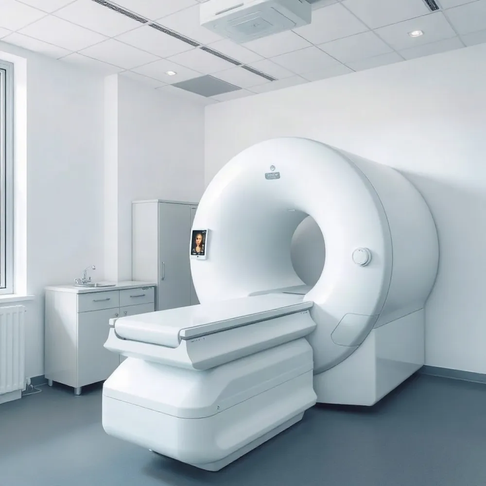
The gold standard for measuring bone mineral density is the dual-energy X-ray absorptiometry (DXA) scan. This quick, painless imaging test focuses on the hip and spine, the two areas most prone to osteoporotic fractures. Healthcare providers typically recommend a baseline DXA around menopause, with follow-up scans every one to two years depending on your results.
DXA Scan. The preferred test for diagnosing osteoporosis, delivering a T-score that compares your bone density to that of a healthy young adult. See more on DXA in postmenopausal women.Peripheral DXA. A portable device that measures bone density in the wrist, heel, or finger. Useful for screening, but results may not fully reflect hip and spine health, so it cannot replace a central DXA.FRAX Tool. A computerized algorithm that estimates your 10-year probability of a major osteoporotic fracture, combining your DXA T-score with clinical risk factors like age, weight, and prior fractures.Quantitative Ultrasound. Uses sound waves rather than radiation to assess bone density, typically at the heel. It is radiation-free and convenient, but considered less precise than DXA for diagnosis.
Many women are surprised to learn that a standard bone density test is not the same as a bone scan performed for other conditions like cancer or infection. A DXA is specifically designed to evaluate fracture risk and is the cornerstone of postmenopausal osteoporosis screening. Professional guidelines from The Menopause Society emphasize early detection, especially since bone loss accelerates rapidly in the years immediately after menopause.
Your healthcare provider may also order additional blood tests to rule out secondary causes of bone loss, such as vitamin D deficiency or thyroid disorders. Understanding which test you need and what the results mean can help you make informed decisions about your bone health plan. Resources like preventing osteoporosis after menopause can guide you through the screening process.

.png)
%201.png)
Compassionate care for every stage of womanhood. Visit one of our convenient locations or book your appointment online today.